Birkedal R, Laasmaa M, Vendelin M
Front Physiol 2014;5:376
PMID: 25324784
Full text: http://dx.doi.org/10.3389/fphys.2014.00376
Abstract
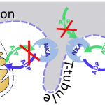
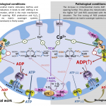
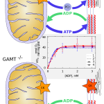
The heart relies on accurate regulation of mitochondrial energy supply to match energy demand. The main regulators are Ca(2+) and feedback of ADP and Pi. Regulation via feedback has intrigued for decades. First, the heart exhibits a remarkable metabolic stability. Second, diffusion of ADP and other molecules is restricted specifically in heart an...
Read More