Peterson P, Kalda M, Vendelin M
Am. J. Physiol., Cell Physiol. 2013 Mar;304(6):C519-31
PMID: 23255581
Abstract
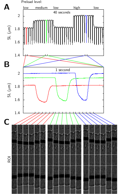
Sarcomere length of a cardiomyocyte is an important control parameter for physiology studies on a single cell level; for instance, its accurate determination in real time is essential for performing single cardiomyocyte contraction experiments. The aim of this work is to develop an efficient and accurate method for estimating a mean sarcomere length of a contracting cardiomyocyte using microscopy images as an input. The novelty in developed method lies in 1) using unbiased measure of similarities to eliminate systematic errors from conventional autocorrelation function (ACF)-based methods when applied to region of interest of an image, 2) using a semianalytical, seminumerical approach for evaluating the similarity measure to take into account spatial dependence of neighboring image pixels, and 3) using a detrend algorithm to extract the sarcomere striation pattern content from the microscopy images. The developed sarcomere length estimation procedure has superior computational efficiency and estimation accuracy compared with the conventional ACF and spectral analysis-based methods using fast Fourier transform. As shown by analyzing synthetic images with the known periodicity, the estimates obtained by the developed method are more accurate at the subpixel level than ones obtained using ACF analysis. When applied in practice on rat cardiomyocytes, our method was found to be robust to the choice of the region of interest that may 1) include projections of carbon fibers and nucleus, 2) have uneven background, and 3) be slightly disoriented with respect to average direction of sarcomere striation pattern. The developed method is implemented in open-source software.