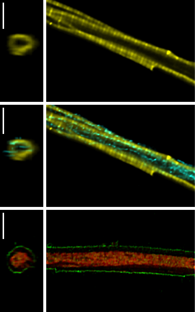
Morphology of trout cardiomyocytes. Confocal microscopy images showing the relatively simple morphology of trout cardiomyocytes. The right image shows one image from a confocal z-stack and the left shows re-constructed cross section. From top to bottom: the first image shows the actin filaments labelled with Atto633-phalloidin (yellow). In second image the same cell as in first, showing also the immunolabelling of tubulin (cyan). In third image, sarcolemma and mitochondria were labelled with Di-8-ANEPPS (green) and Mitotracker Red (red), respectively. Bars are 5 µm.